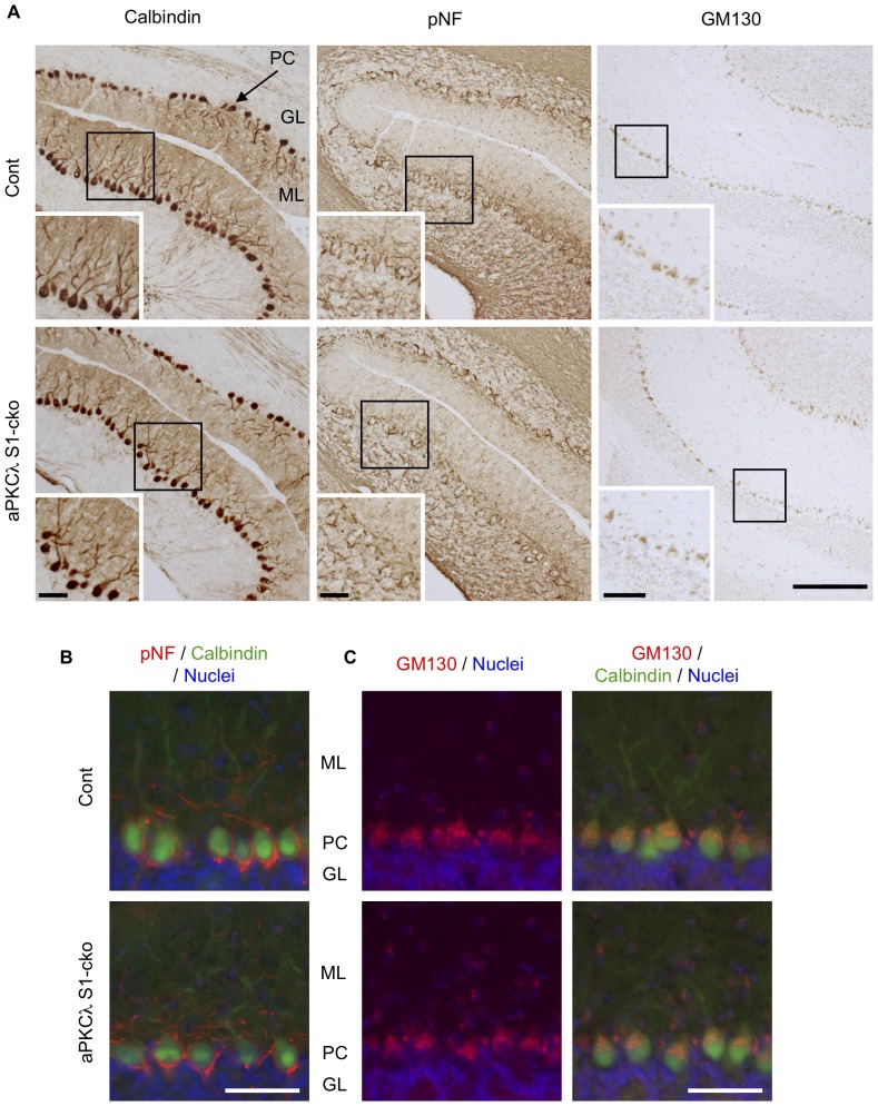
Figure 10. Neural marker staining of aPKCλ deletion mouse cerebellum.
(A) Coronal sections of cerebellum of 7-month-old aPKCλ flox/−; S1-cre (S1-cko) or flox/+ (Cont) female mice were stained with antibodies for calbindin, phospho-neurofilament (pNF) and GM130, markers for Purkinje cells, axons and Golgi apparatus, respectively. Insets are enlarged images of boxed regions. (B, C) The sections were stained with calbindin (green) together with pNF (red; B) or GM130 (red; C). Nuclei were stained with TOTO-3. GM130 was relatively concentrated to molecular layer side in Purkinje cells, whereas pNF was highly detected in granular layer. PC (Purkinje cell), ML (molecular layer) and GL (granular layer). Bars are 200 µm (A) and 50 µm (insets of A, B, C).

