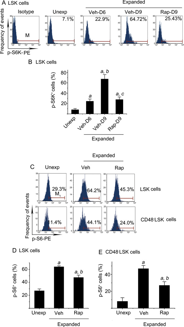
Figure 1. Ex vivo expansion of HSCs activates mTOR in a time-dependent manner.
(A) Representative flow cytometric analyses of phosphorylated S6K (p-S6K) in freshly isolated unexpanded Lin−Sca1+c-kit+ cells (LSK cells) (Unexp) and ex vivo expanded LSK cells after 6-day (Veh-D6) and 9-day (Veh-D9) culture with vehicle (0.1% DMSO) or 9-day (Rap-D9) culture with rapamycin (20 ng/ml) started on day 6 are shown. Cells stained with an isotype control antibody (Isotype) were included as a control for the staining. (B) Percentages of p-S6K+ LSK cells from three independent assays are presented as mean ± SE. a, p<0.05 vs. unexpanded cells; b, p<0.05 vs. cells expanded with vehicle for 6 days; c, p<0.05 vs. cells expanded with vehicle for 9 days. (C) Representative flow cytometric analyses of phosphorylated S6 (p-S6) in freshly isolated LSK and CD48−LSK cells (Unexp) and ex vivo expanded LSK and CD48−LSK cells after 9-day culture with vehicle (Veh, 0.1% DMSO) or rapamycin (Rap, 20 ng/ml) started on day 6 are shown. (D) Percentages of p-S6+ LSK cells from three independent assays are presented as mean ± SE. (E) Percentages of p-S6+ CD48−LSK cells from three independent assays are presented as mean ± SE. a, p<0.05 vs. unexpanded cells; and b, p<0.05 vs. cells expanded with vehicle.

