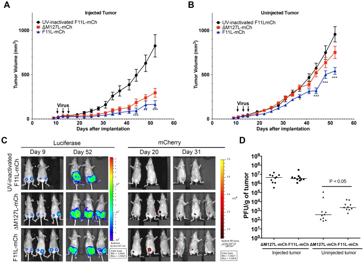
Figure 5. Effect of MYXV F11L-mCh on the growth of secondary untreated tumors.
Two tumors (in opposite mammary fat pads) were established in NIH-III mice using MDA-MB-231 cells. Once palpable, the right-side tumor was injected with three doses of 5x10⁷pfu of ΔM127L-mCh (n = 10), F11L-mCh (n = 10), or an equivalent amount of UV-inactivated F11L-mCh virus (n = 8), and the tumor growth was monitored using calipers, with periodic imaging. After 6 weeks (experiment day 52) the mice were euthanized and the tissues titered to assay for virus. (A) Growth of tumors injected with MYXV. Calipers were used to monitor the growth of the tumors injected with the indicated viruses. (B) Growth of the uninjected contralateral tumor. In both (A) and (B) we report the mean tumor volume ± S.E.M and the statistics compare the cohorts treated with live ΔM127L-mCh or F11L-mCh viruses. (C) Tumor and virus imaging. An IVIS imager was used to detect tumor-encoded luciferase and virus- encoded mCherry on the days indicated. (D) Virus titers in excised tumors. Tumour tissues were recovered from the mice at the experimental endpoint, weighed, and the viruses titered on BGMK cells. The horizontal line denotes the mean titer.

