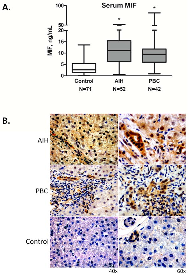
Figure 2.
A. Median serum MIF levels in healthy controls and patients with AIH and PBC (AIH: 11.1±9.55 ng/mL, PBC: 9.58±12.12 ng/mL, Controls: 2.63±3.45 ng/mL, *p<0.001 for AIH vs. Controls and for PBC vs. Controls). There was no significant difference between serum MIF among the two patient cohorts (p=NS). The bottom, middle, and top lines of the box demarcate the 25th, 50th, and 75th percentiles, respectively, and the vertical lines show the maximum and minimum values. B. Immunohistochemistry staining of MIF in human liver tissue from a patient with AIH, a patient with PBC, and a healthy control. The AIH section displays dark MIF staining (dark brown) in 100% of counted hepatocytes, while the PBC section has lighter MIF staining in 100% of counted hepatocytes, with more prominent biliary staining. There was no hepatocyte staining in control tissue. All three sections revealed mild biliary epithelial staining. Images shown are representative of 11 patients examined (AIH=2, PBC=4, Control=5).

