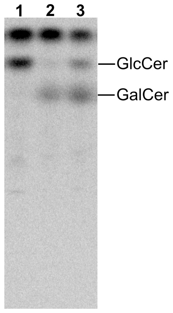
Figure 6. HPTLC pattern of 14C-serine-labelled neutral glycosphingolipids of the MDA-MB-231 cells after doxorubicin treatment.

Cells were grown in medium containing 2 µC/ml 14C-serine for 3 h. Neutral glycosphingolipids were separated in the solvent system 2-propanol/15 M ammonia/methyl acetate/water 75/10/5/15 by vol. The plate was exposed to radiographic screen (DuPoint) for 5 days. The positions of GlcCer and GalCer standards are indicated on the right. Lane 1 – MDA-MB-231 cells grown in complete α-MEM, lane 2 - MDA-MB-231 cells grown in the presence of PPMP for 96 h, lane 3 – MDA-MB-231 cells grown at 90–100% confluence in the presence of 2.5 µM doxorubicin for 48 h.
