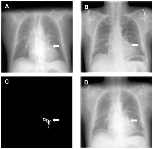
Figure 4.
Visualization of registered CT projection image and dual-energy digital radiography (DR) images. A: Registered CT projection image; B: Dual-energy DR image of the same patient; C: CT projection image of the calcified coronary artery; D: fusion of the three images where the susceptive calcification in the DR image B was verified by the registered CT projection images A and C.

