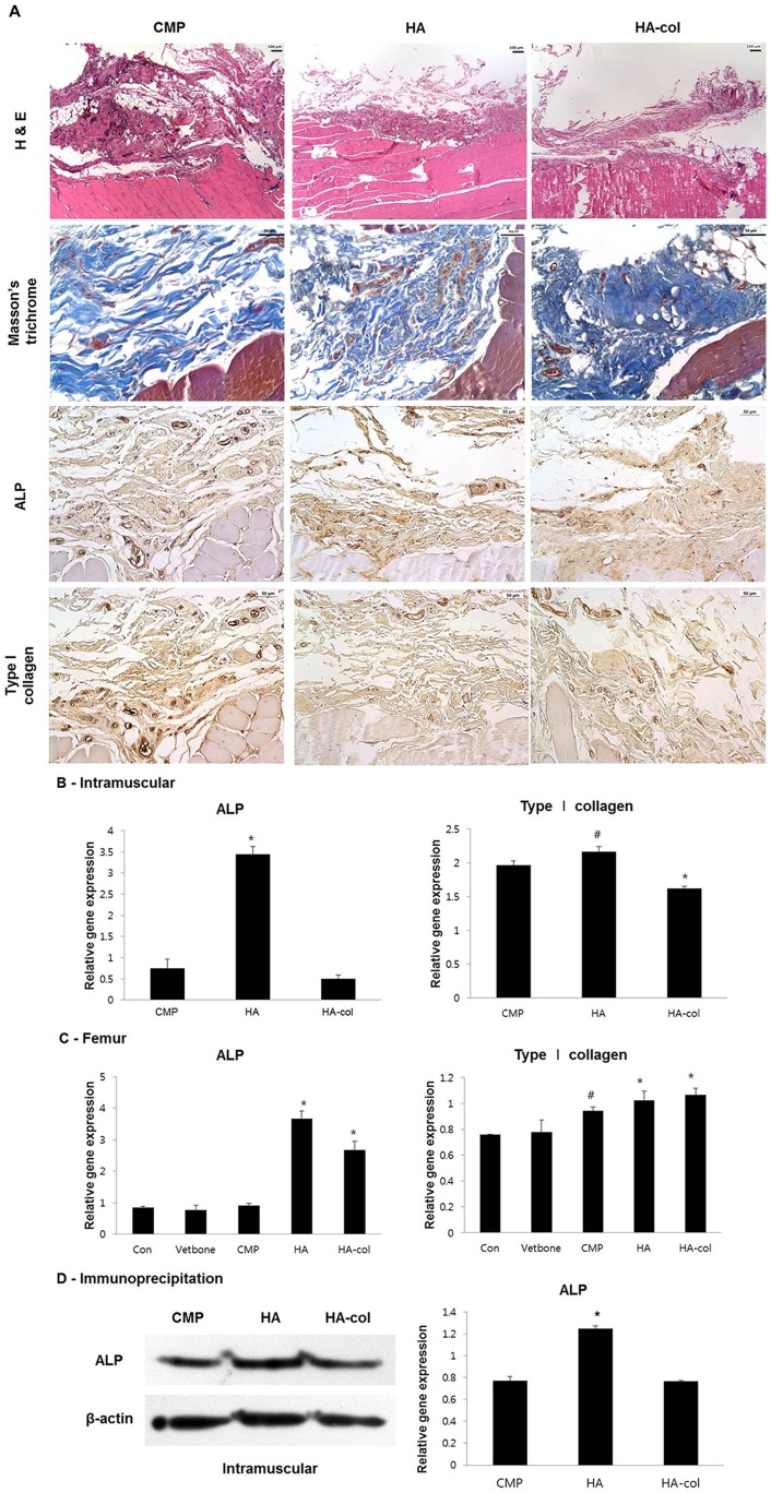
Figure 7. Histological analysis at 12 weeks post-implantation intramuscularly (A).
In H&E staining, peripheral regions of the bioceramic-implanted muscles showed variable degrees of newly formed eosinophilic connective tissue deposition, which show a positive reaction to trichrome staining indicating a collagen-rich stroma. All bioceramic-implanted groups showed positive immunohistochemistry reaction to ALP and type I collagen but differences of positive intensity between each group were not obvious. Immunohistochemistry, Mayer's hematoxylin counter staining, Magnification, ×200. RT-PCR (B, C) for gene and immunoblotting for protein (D) expression of osteogenic molecules in the intramuscularis and cortical defects. (B, C) Formalin fixed paraffin-embedded (FFPE) muscular and femur tissue RNA was extracted by All prep DNA/RNA FFPE kit. In muscles, mRNA expression of ALP was increased to a greater degree in the HA group than in others, with similar results observed by immunoblotting as shown in Figure 6B. Type I collagen was up-regulated in the CMP and HA groups. (D) Expression of ALP was higher in the HA group than in the others, however, there was no significant difference in the expression level of type I collagen among all groups. Protein extraction from FFPE muscular tissues by Q proteome FFPE tissue kit for immunoblotting. Values are the mean ± standard deviation. #p<0.05, *p<0.01 versus the CMP group.

