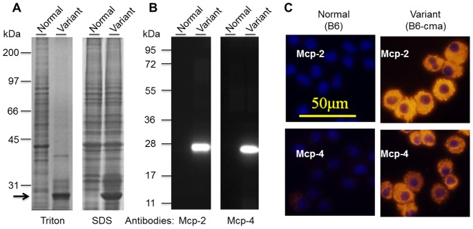
Figure 1. Identification of markedly increased expressions of Mcp-2 and Mcp-4 in BMMCs from a subpopulation of JAK2V617F transgenic mice.

A. Detection of a predominant protein band in cell extracts of BMMCs from a variant line of mice. BMMCs from two JAK2V617F transgenic mice were extracted in a buffer containing 1% Triton X-100 or 1X SDS gel sample buffer were resolved on 10% SDS gel, and proteins were visualized by Coomassie blue staining. The arrow points to a predominant band. B. Verification of Mcp-2 and Mcp-4 over-expressions by Western blotting with specific antibodies. Extracts of BMMCs were separated on 12.5% SDS gel and subjected to Western blotting analyses with anti-Mcp-2 and Mcp-4. C. Verification of Mcp-2 and Mcp-4 over-expression by immunofluorescent cell staining. Mcp-2 and Mcp-4 were probed with specific antibodies followed by Cy-3-conjugated secondary antibodies (red). The nuclei (blue) were revealed by staining with Hoechst 33258.
