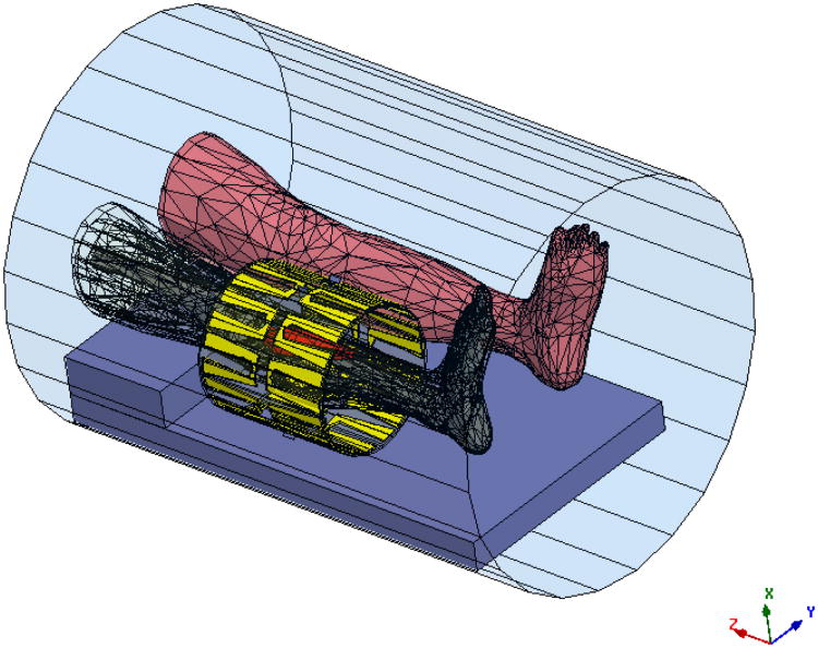
Figure 1.
Electromagnetic segmentation model of the leg as derived from patient CT scan showing meshes for anatomical structures (the red object is tumor) inside 10-antenna cylindrical array. The whole system sits inside the MRI bore. The antennas are evenly separated by 36° around a cylindrical surface of diameter = 23 cm and length =24 cm. The tumor is in the upper right position, next to the upper portion of the tibia, and occupies a volume of about 138.59 cm3. The size of the tumor is 18 cm × 5.65 cm (maximum width).

