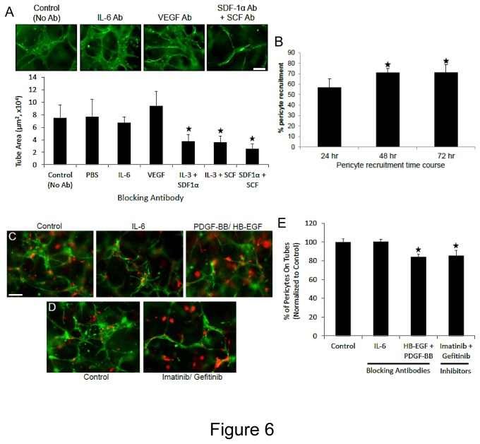
Figure 6. Neutralizing antibodies directed to the hematopoietic cytokines, SCF, IL-3 and SDF-1α markedly block EC tube formation, while antibodies to PDGF-BB and HB-EGF interfere with pericyte assembly on EC-lined tubes in 3D fibrin matrices under serum-free defined conditions.
GFP-ECs were primed with hematopoietic cytokines (factors) or VEGF/FGF-2 overnight and then were seeded into fibrin matrices with hematopoietic factors and FGF-2 in the presence of Cherry-pericytes and in the presence of the indicated neutralizing antibodies or chemical inhibitors. (A) The antibodies were added at 50 µg/ml (IL-6, SCF, IL-3, SDF-1α) or 100 µg/ml (VEGF) versus controls and after 72 hr, cultures were fixed, photographed and quantitated for EC tube formation (n=10, values are averaged + SD, asterisks indicate significance compared to control, p<.01). (B) Time course of pericyte recruitment to EC tubes using real-time movies. At the indicated times, the percentage of pericytes that were associated with EC tubes were quantitated (n=8, values are averaged + SD, asterisks indicate significance compared to control, p<.01). (C,D) EC-pericyte co-cultures were established in the absence or presence of the indicated blocking antibodies (each added at 50 µg/ml) or chemical inhibitors (each added at 1 µM). After 72 hr, cultures were fixed and photographed (C,D) or were quantitated for pericyte recruitment (E) (n=6, values are averaged + SD, asterisks indicate significance compared to control, p<.01). Bar equals 100 µm.

