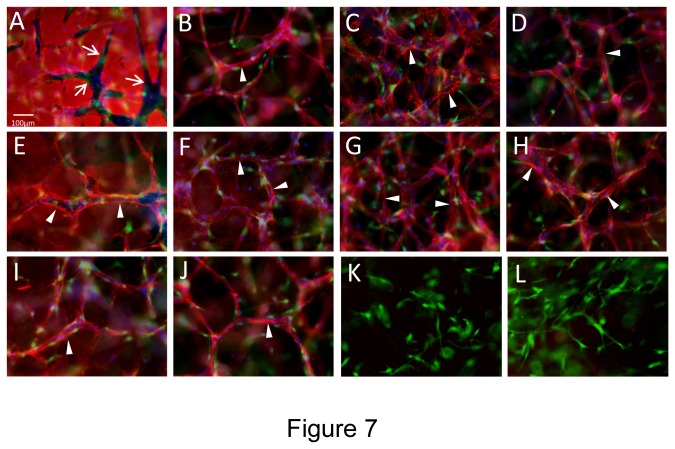
Figure 7. Hematopoietic stem cell cytokines stimulate EC-pericyte tube co-assembly and vascular basement membrane matrix deposition under serum-free defined conditions in 3D fibrin matrices.
ECs were primed with VEGF/FGF-2 overnight and then were seeded with GFP-pericytes into 3D fibrin matrices containing SCF, IL-3, SDF-1α, Flt-3L, and FGF-2. After 120 hr, cultures were fixed and stained for extracellular antigens using various antibodies directed to: (A) Fibrin; (B) CD31; (C) Collagen type IV; (D) Laminin; (E) Fibronectin; (F) Perlecan; (G) Nidogen-1; (H) Nidogen-2; (K) Collagen type I; and (L) Collagen type III. In some cultures, the fibrin gel was supplemented with bovine fibronectin (I, J) and a monoclonal antibody that is specific for human fibronectin was used to assess fibronectin deposition within the vascular basement membrane (I). In this case, CD31 antibody staining was shown for comparison (J). In all other cases, the fibrin gels were supplemented with human fibronectin (A-H, K,L). Arrows indicate the borders of vascular guidance tunnels which are generated during EC tubulogenesis and both ECs and pericytes reside within these tunnel spaces (A). Arrowheads indicate vascular basement membrane deposition. Bar equals 25 µm.

