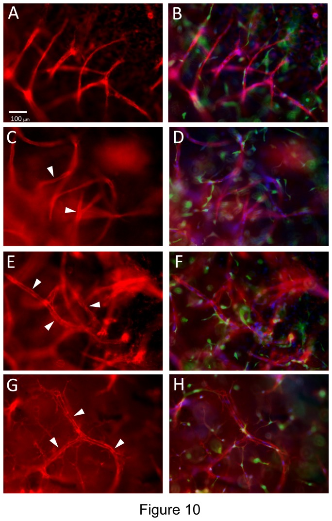
Figure 10. Pericytes recruit to invading EC sprouts/tubes and induce vascular basement membrane assembly on these EC tubes in 3D fibrin matrices under serum-free defined conditions.
GFP-pericytes were seeded into 3D fibrin matrices which contained SCF, IL-3, SDF-1α, Flt-3L and FGF-2 while ECs were primed with VEGF/FGF-2 and then seeded on the surface of polymerized fibrin gels that contained growth factors and pericytes. ECs were allowed to sprout and form tubes for 120 hr prior to fixation and immunofluorescence staining with antibodies to CD31 (A,B); laminin (C,D); collagen type IV (E,F); or fibronectin (G,H). The basement membrane matrix antigens were stained in the absence of detergent to examine only extracellular deposition of these molecules. The right sided images (B,D,F,H) are overlay images for each stain showing the antigen stain in red, while GFP-pericytes are green and nuclei are blue (stained with Hoechst dye). Arrowheads indicate vascular basement membrane matrix deposition. Bar equals 100 µm.

