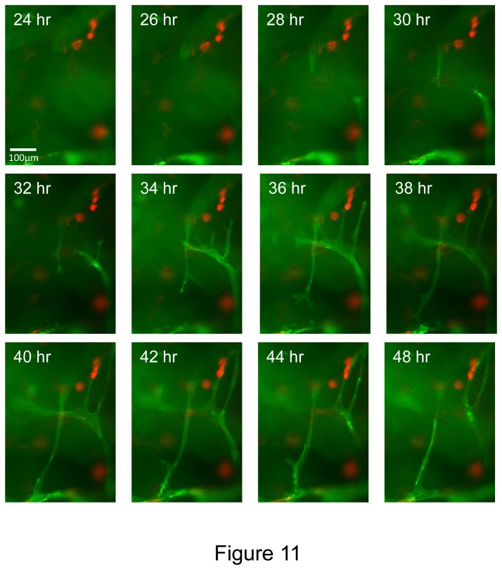
Figure 11. Time lapse images of EC sprouting and tubulogenesis in 3D fibrin matrices under serum-free defined conditions which are stimulated by hematopoietic stem cell cytokines, FGF-2 and pericytes.
mCherry-pericytes, hematopoietic cytokines and FGF-2 were incorporated into the fibrin matrix, and VEGF/FGF-2 primed ECs (membrane AcGFP-labeled) were seeded onto the surface of fibrin gels and real-time movies were made. Select time points are shown from a representative field showing marked EC sprouting and tube formation as well as EC-pericyte interactions. Interestingly, it appears that EC sprouts might be preferentially orienting toward mCherry-pericytes in these movies suggesting the presence of pericyte-derived directional cues. Bar equals 100 µm.

