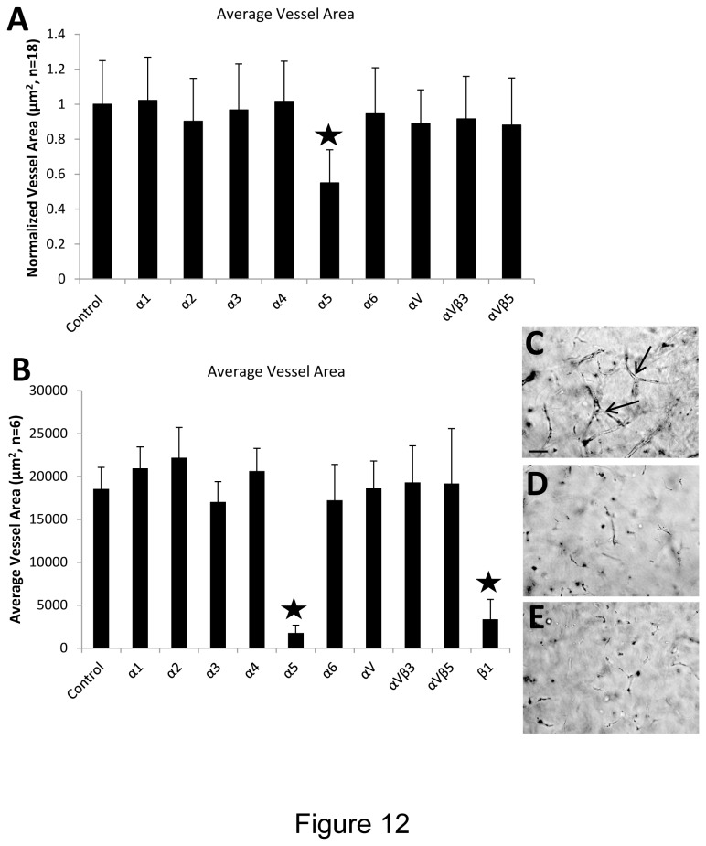
Figure 12. Integrin α5β1 controls EC sprouting and tubulogenesis in 3D fibrin matrices under serum-free defined conditions.
(A) EC-pericyte co-cultures were established by suspending both cell types into fibrin matrices along with hematopoietic cytokines and FGF-2. The indicated anti-integrin blocking antibodies were added at 20 µg/ml and after 72 hr, EC tube area was determined using Metamorph software. Asterisk indicates significance at p< .01 compared to the control condition (n=18, values are averaged + SD). (B) EC sprouting assays were established by seeding ECs on the surface of fibrin gels which contained pericytes as well as hematopoietic cytokines and FGF-2. The indicated anti-integrin blocking antibodies were added at 20 µg/ml and after 72 hr, EC tube area in the invading sprouts was quantitated using Metamorph software. Asterisks indicate significance at p< .01 compared to the control condition (n=6, values are averaged + SD). Representative photographs of invading sprouts and EC tubulogenesis in the control condition (C); anti-α5 integrin subunit (D); and anti-β1 integrin subunit (E). Bar equals 100 µm.

