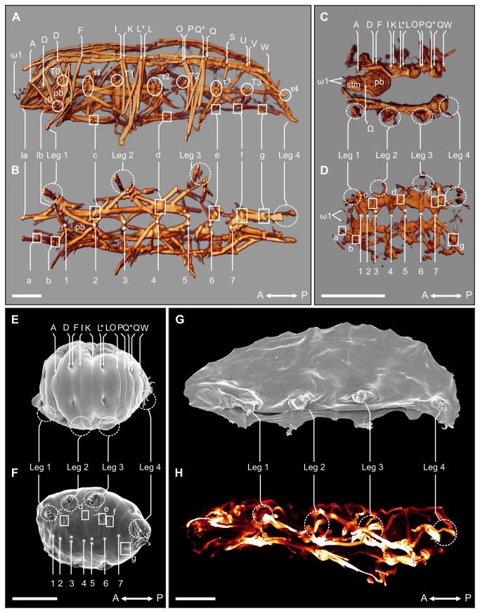
Figure 3. Myoanatomical changes in Richtersius coronifer during tun formation.
A-D. 3D reconstructions of the tardigrade musculature as visualized by fluorescent phalloidin. A. Lateral view of active (hydrated) state showing details of the dorsal, lateral and leg musculature. B. Ventral view showing details of the ventral longitudinal musculature in the active state. C. Dorsal view of the myoanatomy of the tun (dehydrated) state D. Ventral view of the myoanatomy of the tun. E-F. Scanning electron microscopy (SEM) of animals in the tun state showing the corresponding external morphology of the myoanatomy presented in C and D. E. Dorsal view of the tun. F. Ventral view of the tun. G. SEM of an animal incubated in 1.0 mg/ml phalloidin for 24 h and subsequently dehydrated. The animal failed to form a tun upon dehydration, and collapsed into a flattened shape. A similar collapse was seen in DNP exposed animals upon dehydration. H. Corresponding maximum projection image of a confocal z-series of the musculature of a specimen incubated in 1.0 mg/ml phalloidin for 24 h before dehydration. A↔P, anterior-posterior axis; A-W, dorsal attachment sites; τ0-τ4, lateral attachment sites; la-g, ventral intermediate attachment sites; 1-7, ventromedian attachment sites; pb, pharyngeal bulb; ω1-Ω, attachment sites of muscles associated with the pharyngeal bulb; stm, stylet muscles. Solid circles indicate lateral attachment sites, solid squares show ventral intermediate attachment sites, while dashed circles indicate areas of the legs. Scale bars = 100 μm.

