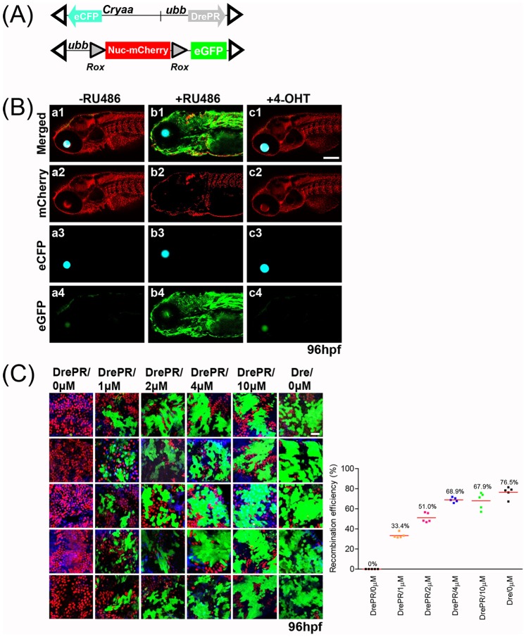
Figure 2. Induction of DrePR recombinase activity by RU486.
(A) Schematic of ubb-DrePR driver line and Rox-Nuc-mCherry-Rox reporter. Additional cryaa:eCFP cassette facilitates identification of transgene-expressing embryos. Open triangles indicate Tol2 arms. (B) Tight control of DrePR recombinase activity by RU486. ubb-DrePR; Rox-Nuc-mCherry-Rox-eGFP embryos were treated with and without 4 µM RU486 between 24 and 48 hpf, and imaged at 96 hpf. No expression of eGFP is observed in untreated (-RU486) or tamoxifen (4-OHT)-treated embryos, while treatment with RU486 results in potent induction of eGFP expression indicating successful recombination of Rox-Nuc-mCherry-Rox-eGFP allele. (C) To quantify the efficiency of Dre recombination in Dre-expressing embryos and DrePR recombination in DrePR-expressing embryos, the intestine of larval zebrafish (4 dpf) were dissected following treatment with RU486 at the indicated concentration between 24–48 hpf. DAPI-labeled cells also labeled by either nucleus mCherry or cytoplasmic eGFP were counted. Maximal recombination frequency is achieved an an RU486 concentration of 4 µM, at a level comparable with Dre lacking the PR fusion. Scale bar: 25 µm.

