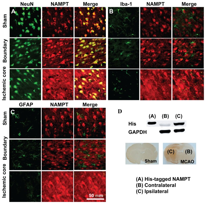
Figure 2. Distribution of NAMPT protein in rat brain after MCAO.
(A-C) Representative images of double immunofluorescent staining of NeuN (for neuron) and NAMPT (A), Iba-1 (for microglia) and NAMPT (B), GFAP (for astrocyte) and NAMPT (C) in rat brain after 30 min MCAO and 24 h reperfusion. Immunofluorescent staining was repeated on three rats. (D) Using Western blot (upper panel) and immunochemistry staining (lower panel), the i.v. injected His-tagged NAMPT was detected in the ischemic brain area after 30 min MCAO and 24 h reperfusion.

