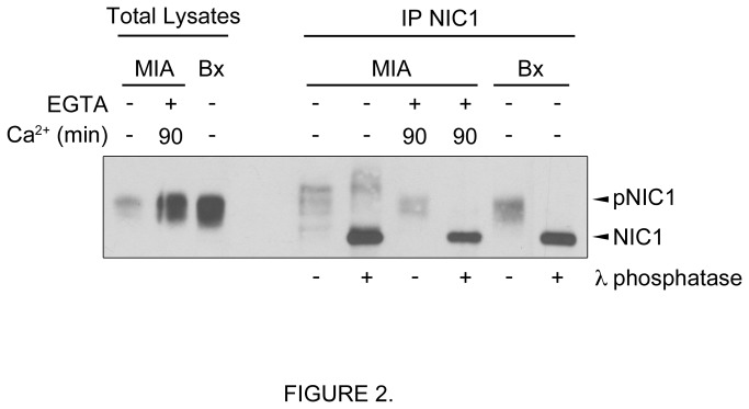
Figure 2. NIC1 is phosphorylated in pancreatic cancer cells.
NIC1 was immunoprecipitated (IP NIC1) from total lysates of exponentially growing MIA PaCa-2 (MIA), MIA PaCa-2 cells treated with EGTA (4mM) for 15min and then returned to their normal culture media for 90min, or exponentially growing BxPC-3 (Bx). Following NIC1 immunoprecipitation, phosphatase assays were performed (+) using the lambda phosphatase (λ phosphatase). The IP NIC1 not incubated with the lambda phosphatase is indicated as pNIC1. A representative Western blot using an antibody against NIC1 is shown.

