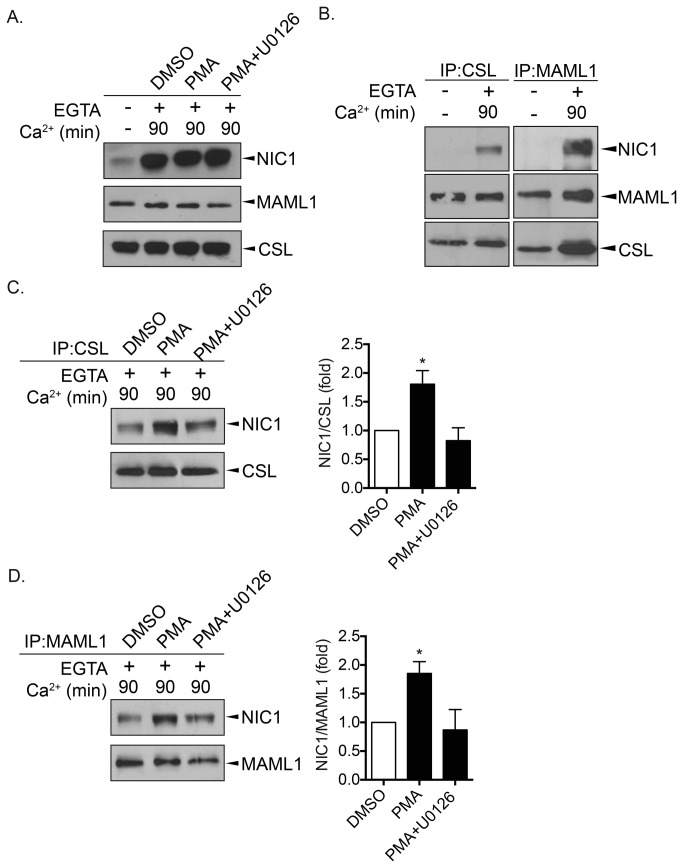
Figure 5. Activation of the MEK/ERK pathway promotes NIC1 association with its transcriptional partners.
MIA PaCa-2 cells were left untreated (-) or treated with EGTA (4mM) for 15min (+). EGTA-containing medium was removed and replaced by normal culture media (Ca2+) containing DMSO, PMA (100nM) or PMA + U0126 (10μM) for 90min. A. NIC1, MAML1 and CSL expression levels were assessed by western blot using the appropriate antibodies. B. CSL and MAML1 were immunoprecipitated (IP) prior to western blot analyses using the indicated antibodies. C. CSL was immunoprecipitated (IP) followed by western blot analyses of NIC1 (left). A graphic representation depicting the relative amount of NIC1 co-immunoprecipitated with CSL (NIC1/CSL) is shown (right). D. MAML1 was immunoprecipitated (IP) followed by western blot analyses of NIC1 (left). A graphic representation depicting the relative amount of NIC1 co-immunoprecipitated with MAML1 (NIC1/MAML1) is shown (right). C. and D. The results are the means ± SEM of at least three independent experiments. * p<0.05 compared to DMSO-treated cells.

