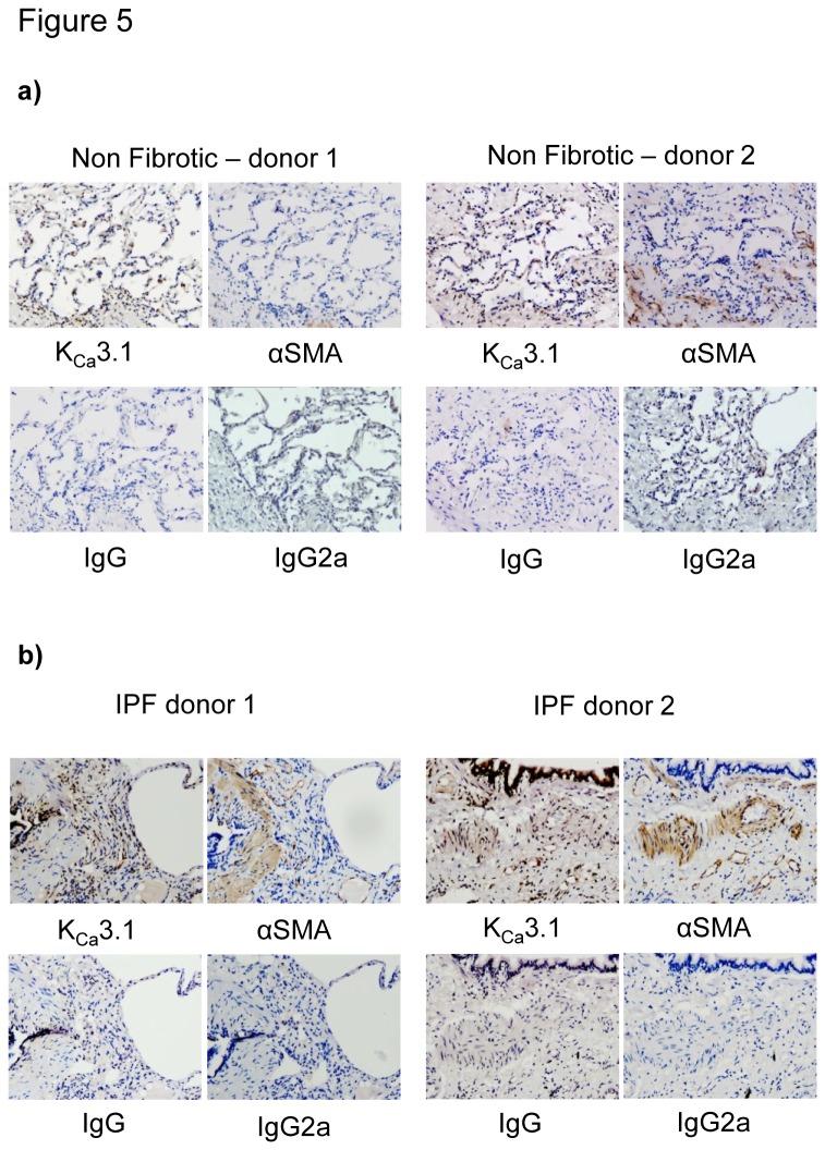
Figure 5. KCa3.1 expression within human lung tissue of non-fibrotic and IPF patients.
a) Representative KCa3.1 and αSMA immunostaining of healthy lung parenchyma from two NFC tissue donors. All pictures are from sequential sections. Isotype controls are negative. b) Representative immunostaining of lung parenchyma from two IPF tissue donors demonstrating KCa3.1 and αSMA immunostaining in areas of fibrosis. All pictures are from sequential sections. KCa3.1 channel expression is particularly strong in the epithelium and within and surrounding areas positive for αSMA.

