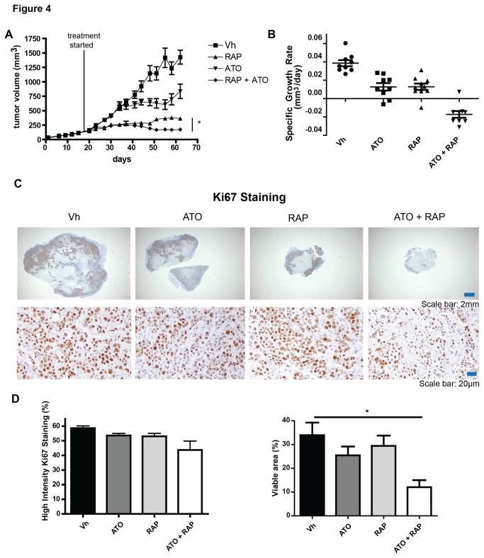
Figure 4. Combination treatment of ATO and rapamycin significantly inhibits tumor growth in an MDA-MB-468 xenograft model.
Mice were implanted with MDA-MB-468 cells in the mammary fat pad and once tumors reached 100-200 mm3, mice were treated with vehicle (Vh; 20% PEG 400 and 20% TWEEN 80), 7.5mg/kg rapamycin, 7.5 mg/kg ATO, or rapamycin plus ATO every other day. Tumor size was monitored (A) and specific growth rate of the tumor (B) was calculated from days 37-62 and is expressed as change in tumor volume in mm3/day. Data are expressed as mean with standard error bars (n = 8-9 mice per group). Tumors were stained with antibodies detecting the proliferative marker Ki67. Representative pictures at low and high magnification of each treatment group are shown (C). Percent cells with high intensity staining and positive Ki67 staining per tumor area were calculated using algorithms provided with the ImageScope software. * = p<0.05 Tumors from individual mice are represented with mean and standard error bars (n = 4-5 mice).

