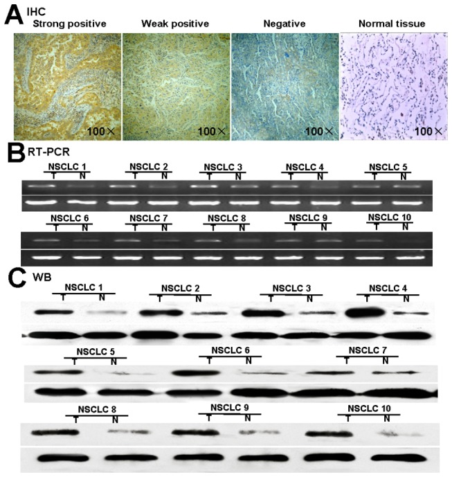
Figure 2. Expression of DKK1 in NSCLC tissues.

(A) Expression by immunohistochemical staining. DKK1 strong positive expression in lung cancer showing staining mainly in the cytoplasm of tumor Cells (magnification 100×). Representative DKK1 weak positive lung cancer with pale yellow particles in tumor cells (magnification 100×). DKK1 negative lung cancer cell showing almost no appreciable staining of tumor cells (magnification 100×). Normal lung tissue without staining. (B) Expression of DKK1 mRNA in NSCLC tissues and matched normal lung tissues. (C) Expression of DKK1 protein in NSCLC tissues and matched normal lung tissues. N, Normal tissue; T, Tumor tissue. Positive expressions of DKK1 were mainly accompanied with lymph nodes metastasis.
