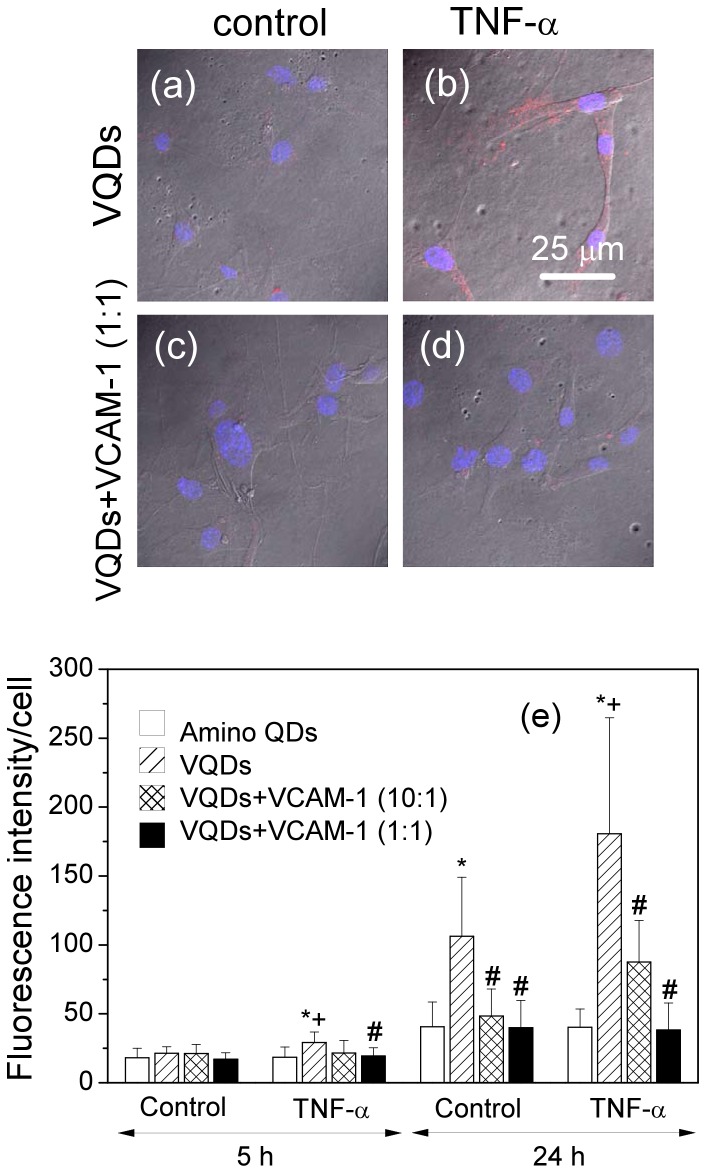
Figure 4. Conjugation with VCAM-1 binding peptide specifically increases the QD fluorescence signal in the VCAM-1 expressing endothelial cells.
Confocal images of mouse endothelial cells: (a) Control cells incubated with VCAM-1 binding peptide functionalized QDs (VQDs) for 24 h; (b) TNF- treated cells incubated with VQDs for 24 h; (c) Control cells incubated with pre-incubated mixture of VQDs and recombinant VCAM-1 (1∶1) for 24 h; (d) TNF-
 treated cells incubated with pre-incubated mixture of VQDs and recombinant VCAM-1 (1∶1) for 24 h. Blue signal comes from DAPI nuclei staining and red signal from QDs. (e) Quantification of fluorescence intensity. Values are means
 SD.
SD.  vs QD within the same treatment group and time point;
  vs control VQD within the same time point;
  vs VQD within the same treatment and time point.

