Figure 1.
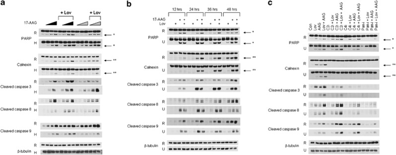
Simultaneous inhibition of HSP90 and the IBP results in the induction of apoptosis of myeloma cells. Immunoblots depicting PARP, calnexin, cleaved caspase 3, cleaved caspase 8, cleaved caspase 9 and β-tubulin are shown. * denotes the PARP cleavage product while ** denotes the calnexin cleavage product. β-Tubulin is used as a loading control. Data are representative of at least three independent experiments. (a) RPMI-8226 (R) and H929 (H) cells were incubated with lovastatin (Lov, 10 μM for RPMI-826 and 2.5 μM for H929) and/or varying concentrations of 17-AAG (AAG) for a total of 48 h. The wedges indicate increasing concentrations of 17-AAG (0.1, 0.5, 1 μM for RPMI-8226 cells and 0.05, 0.1, 0.25 μM for H929 cells). The black wedges indicate that 17-AAG was added at the start of the 48-h incubation, whereas the gray wedges indicate that 17-AAG was added after 24 h. (b) RPMI-8226 (R) and U266 (U) cells were cultured for 12–48 h in the presence or absence of lovastatin (10 μM) and/or 17-AAG (0.5 μM). (c) Effects of caspase inhibitors on 17-AAG- and lovastatin-induced apoptosis. RPMI-8226 and U266 cells were incubated for 48 h in the presence or absence of 17-AAG (0.5 μM), lovastatin (10 μM), caspase-2 inhibitor (50 μM VD-VAD, C2i), caspase-4 inhibitor (50 μM Y-VAD, C4i) or pan-caspase inhibitor (50 μM VAD, Pan-i).

