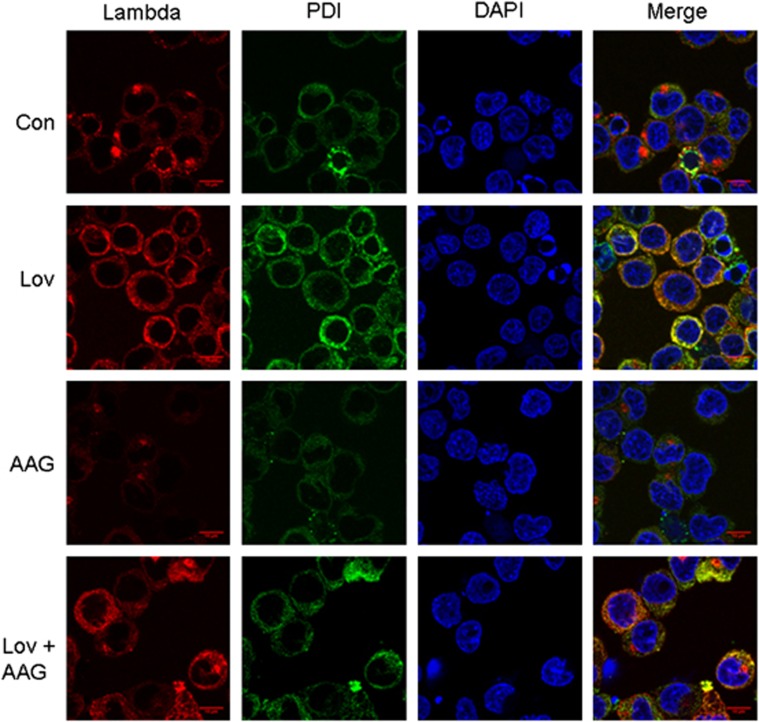
Figure 3.
Changes in intracellular lambda light chain localization following drug treatment. RPMI-8226 cells were incubated in the absence or presence of 10 μM lovastatin (Lov) and/or 0.5 μM 17-AAG (AAG) for 24 h. Indirect immunofluorescence microscopy was performed as described in the Materials and Methods section using antibodies directed against lambda light chain and PDI. DAPI was used for nuclear staining. The merged images represent merging of the lambda, PDI and DAPI images via ImageJ software.

