Figure 7.
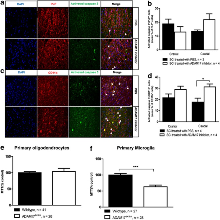
ADAM17 inhibition in vivo or deficiency in vitro increases microglial apoptosis. (a) Representative pictures of PLP/activated caspase-3 double staining. Scale bar=20 μm. The arrows indicate apoptotic oligodendrocytes. (b) Quantification of the number of apoptotic oligodendrocytes (PLP/activated caspase-3 double-positive) in PLP-eCFP transgenic mice in a squared area of 225 × 225 μm around the lesion site. There is a slight but non-significant increase in the number of apoptotic oligodendrocytes caudal to the lesion site (225 μm; n=3-4 mice per condition). (c) Representative pictures of CD11b+/activated caspase-3 caudal to the lesion site. The arrows show apoptotic microglia in both groups. Scale bar=20 μm. (d) A significant increase in the number of apoptotic microglial (CD11b/activated caspase-3 double positive) cells caudal to the lesion site was found after inhibition of ADAM17 (n=4 mice per condition; *P<0.05). (e) Survival of primary immature oligodendrocyte cultures from ADAM17-deficient mice is not affected. (f) Primary microglial cultures from ADAM17-deficient mice have a reduced survival compared with microglial cultures from wild-type animals (n=26–41 wells (primary oligodendrocytes), n=27–28 wells (primary microglia)(***P<0.001)

