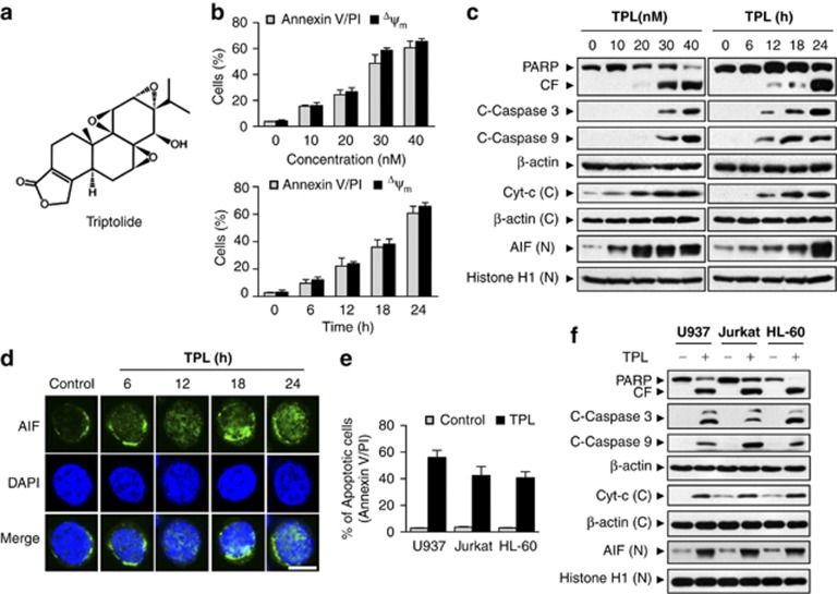
Figure 1.
Triptolide induced apoptosis and mitochondrial injury in multiple leukemia cell lines. (a) The chemical structure of triptolide, C20H24O6, molecular weight: 360.4. (b and c) U937 cells were treated with various triptolide (TPL) concentrations for 24 h or with 40 nM triptolide for different lengths. The percentage of apoptotic cells was determined by FACS analysis using Annexin V/PI staining. Mitochondrial membrane potentials (ΔΨm) were detected by rhodamine-123 staining and flow cytometry. Values represent the mean±S.D. for five separate experiments. Total protein lysates, nuclear extracts, and cytosolic fractions were analyzed by immunoblotting using the indicated antibodies. (d) After triptolide treatment, cells were collected and stained with anti-AIF (green) and 4′,6-diamidino-2-phenylindole (DAPI; blue) to identify cellular nuclei. Fluorescence was visualized by a laser confocal scanning microscope. Scale bar represents 10 μm. These data are representative of three independent experiments. (e and f) U937, Jurkat, and HL-60 cells were treated with or without 40 nM triptolide for 24 h, after which apoptosis was determined by FACS analysis using Annexin V/PI staining. Total protein lysates, nuclear extracts, and cytosolic fractions were analyzed by immunoblotting using the indicated antibodies. CF, cleavage fragment; C, cytosolic fractions; N, nuclear extracts

