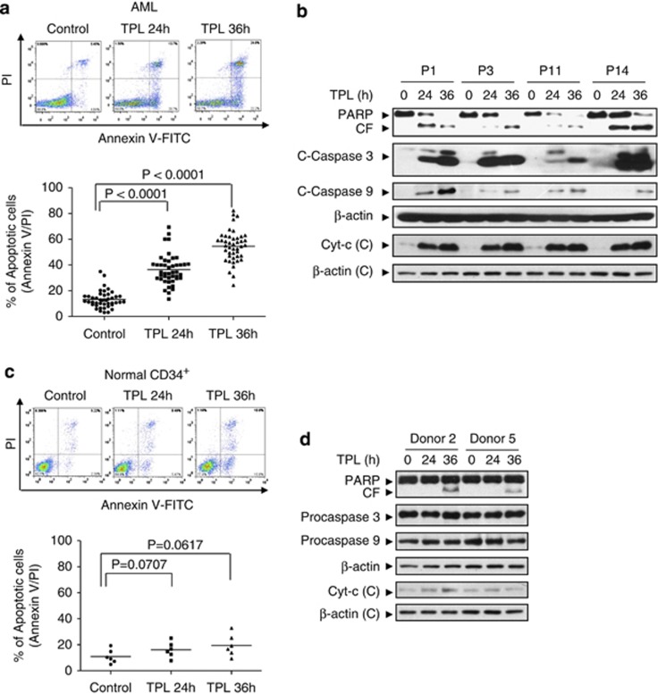
Figure 2.
Triptolide induced apoptosis in primary human leukemia blasts but not in normal CD34+ cells. (a) Primary leukemia blasts from 44 patients were treated with 40 nM triptolide for 24 or 36 h, after which apoptosis was determined by FACS analysis using Annexin V/PI staining. Upper panel: representative FACS images. Lower panel: numbers indicate the percentage of apoptotic cells; each symbol represents results from individual patients. (b) Total protein lysates and cytosolic fractions of the samples from four AML patients were then obtained and analyzed by immunoblotting using the indicated antibodies. (c) Normal CD34+ cells were isolated from 6 normal bone marrow samples and exposed to 40 nM triptolide for 24 or 36 h. The percentage of apoptotic cells was determined by FACS analysis using Annexin V/PI staining. (d) Two normal donors were treated with 40 nM triptolide for 24 or 36 h. Total protein lysates and cytosolic fractions of these samples were then obtained and analyzed by immunoblotting using the indicated antibodies

