Figure 8.
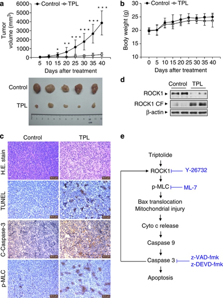
Triptolide inhibited the growth of U937 xenografts. A total of 24 nude mice were inoculated with U937 cells and randomly divided into two groups (12 mice/group) that were treated with either vehicle or triptolide. (a) Tumor volumes were measured at the indicated intervals (upper panel) and images are shown for five representative tumors from each group after 40 days of treatment (lower panel). Data are shown as the mean±S.D. *P<0.05; **P<0.01; ***P<0.001. (b) Changes in body weight in the mice during 40 days of triptolide treatment. (c) Tumors were fixed and stained with H&E to examine tumor cell morphology. TUNEL assays were used to determine the apoptotic effects of triptolide. Immunohistochemistry was used to determine the levels of cleaved caspase-3 and phospho-MLC. Scale bar represents 50 μm. (d) After treatment, tumors from the vehicle and triptolide groups were lysed and subjected to immunoblotting with anti-ROCK1. Results are representative of three independent experiments. (e) An illustration of the molecular mechanism of triptolide-induced apoptosis. Exposure to triptolide results in activation of ROCK1 and MLC and MYPT phosphorylation, leading in turn to Bax translocation, culminating in cytochrome c release (Cyto c), caspases activation, and apoptosis. Activated caspase-3 in turn causes activation of ROCK, thus creating a positive feedback loop

