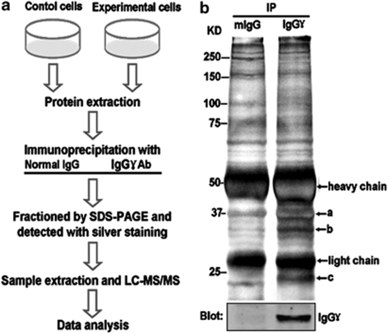
Figure 2.
Identification of cancer-derived IgG-associated proteins. (a) Schematic illustration of the strategy used to screen cancer-derived IgG-associated proteins. (b) Proteins immunoprecipitated with mouse anti-human IgG (γ chain-specific) antibody or normal mouse IgG from total lysates of HeLa cells were fractionated with 10% SDS-PAGE gel. The gels were either visualized with silver staining (upper panel) or blotted with anti-IgGγ antibodies (lower panel). The differential bands (marked a–c) were subjected to trypsin digestion and LC-MS/MS analysis. The identified proteins were listed along with the corresponding bands (Supplementary Table S1)

