Figure 2.
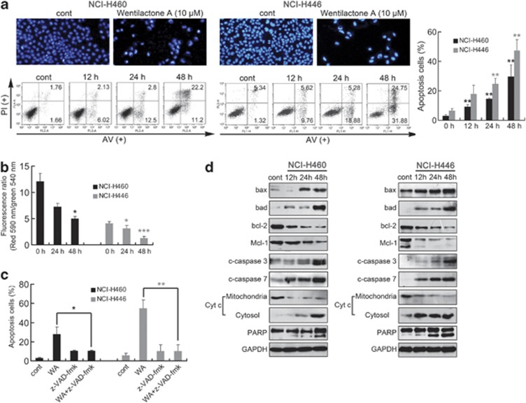
WA triggers mitochondrial-related apoptosis in NCI-H460 and NCI-H446 cells. (a) WA-induced morphological changes as indicated by DAPI staining. Representative photomicrographs of control and 48 h 10 μM WA treatment groups are showed. Apoptosis in NCI-H460 and NCI-H446 cells was assessed after 12–48 h of treatment with 10 μM WA by Annexin V-FITC/PI binding and measured by flow-cytometry analysis. Numbers indicate the percentage of cells in each quadrant. **P<0.01 versus the drug-untreated group. (b) The mitochondrial membrane potential was measured using JC-1 by flow cytometry. Decrease in the ratio of the green fluorescence (FITC) to the red fluorescence (PE) indicates loss of ΔΨm. (c) NCI-H460 and NCI-H446 cells were treated with 20 μM z-VAD-fmk, a caspase inhibitor, for 1 h before treatment with 10 μM WA for 48 h. Apoptosis was assessed by flow cytometry as mentioned above. *P<0.05, **P<0.01. (d) Western blot of cells analyzed for bax, bad, bcl-2, Mcl-1, cleaved caspase-3/7, PARP, cytochrome c in cytosol and mitochondria after treatment with 10 μM WA for the indicated time periods. Results are representative of three separate experiments. GPDH is shown as protein-loading control

