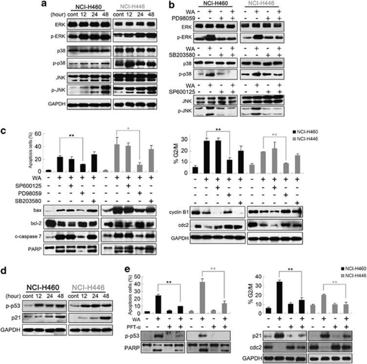
Figure 4.
ERK and p53 regulate WA-induced apoptosis and G2/M arrest. (a) Cells were treated with 10 μM WA for various incubation times, total and phosphorylated MAPK members (JNK, p38, ERK) were detected using immunoblot assay. (b) Cells were pre-treated with 20 μM MEK inhibitor (PD98059), JNK inhibitor (SP600125) or p38 inhibitor (SB203580) followed by treatment with or without 10 μM WA for 48 h. Total and activation forms of ERK, JNK or p38 were evaluated by western blotting; (c) Cells were pre-treated with the indicated inhibitor for 1 h, then treated with 10 μM WA for 48 h and analyzed for apoptosis, cell-cycle analysis and expression of bax, bcl-2, cleaved caspase-7, PARP, cyclin B1 and cdc2. (d) p-p53 (Ser 15) and p21 protein levels were detected after WA treatment. (e) Cells were incubated for 1 h in the presence or absence of PFT-α (20 μM) and then 10 μM WA was added followed by incubation for an additional 48 h. The induction of apoptosis and cell-cycle distribution were determined by flow cytometry. Apoptosis and G2/M checkpoint-related proteins were detected by western blot. Values are means±S.D. from three independent experiments. *P<0.05, **P<0.01

