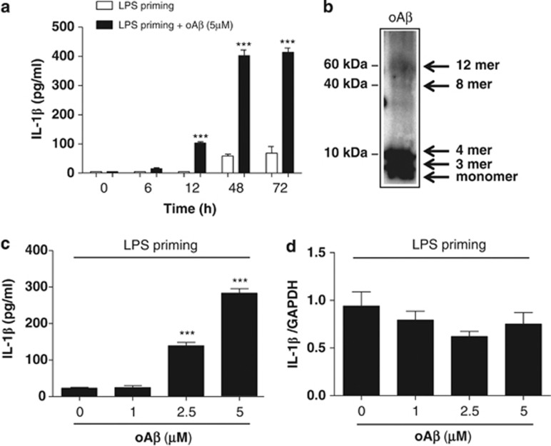
Figure 1.
oAβ induces IL-1β release in LPS-primed microglia. Microglia primed with LPS for 3 h were washed with ice-cold PBS and treated with oAβ for varying times; the concentration of IL-1β in the culture supernatant was then measured (a). ***P<0.001, versus 0 h. Western blot analysis of oAβ used in the present study (b). The blot was incubated with mouse anti oAβ monoclonal antibodies (6E10) (1 : 1000, Chemicon). LPS-primed microglia were treated with oAβ for 48 h, and the concentration of IL-1β in the culture supernatant as well as mRNA expression were measured by ELISA (c) and qPCR (d). Data indicate means±S.D. for five independent experiments. ***P<0.001, versus LPS-primed microglia

