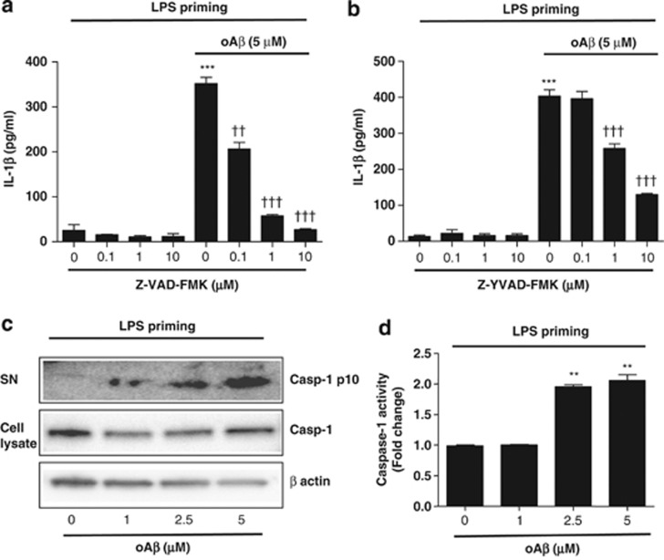
Figure 2.
oAβ induces IL-1β secretion/release via caspase-1 activation. LPS-primed microglia were treated with Z-VAD-FMK (a) or Z-YVAD-FMK (b) for 30 min before oAβ stimulation, and IL-1β in the culture supernatant was measured at 48 h. Data indicate means±S.D. for four independent experiments. ***P<0.001, versus LPS-primed microglia as control (ctl). ††, or ††† denotes P<0.01, or 0.001, respectively, versus LPS-primed microglia+oAβ. (c) After LPS-priming microglia were treated with oAβ for 48 h, Casp-1 p10 in the culture supernatant as well as caspase-1 and β-actin in the cell lysates were assessed by western blotting. Data are representative of two independent experiments. (d) LPS-primed microglia were treated with oAβ for 48 h, and caspase-1 activity was measured. Data indicate means±S.D. for three independent experiments. **P<0.01, versus LPS-primed microglia without oAβ

