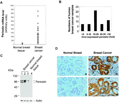
FIG. 1.
High levels of periostin expression are associated with human breast cancers. (A) Periostin expression pattern based on gene array data. The raw data from gene array analysis of the expression of periostin in normal (3 samples) and breast cancer (50 samples) tissues are plotted (the mean value for normal tissues at 106 as a baseline versus that for breast cancer samples at 2,100). (B) The same gene array data on periostin expression by 50 breast cancers were categorized into different groups based on levels of periostin overexpression. (C) Periostin protein expression in normal and tumor tissue samples. Tissue extracts from normal or breast cancer tissue samples were subjected to immunoblot analysis with a polyclonal antiperiostin antibody. The results shown in lanes 1 and 2 are from tumor samples from the group with the highest levels of periostin mRNA expression (>30-fold). The result shown in lane 3 was from tumor tissue with less than a fivefold increase in periostin mRNA expression. The higher band of the doublet is likely the unprocessed form of periostin, and the lower band is likely the secreted form of periostin with its signal sequence cleaved from the N terminus. (D) Tissue sections from human normal breast and breast cancer samples were analyzed by immunohistochemical staining and representative sections of each type of sample are shown. Red immunostaining represents positive staining for periostin protein in tumor tissues but not in normal tissues (magnification, ×200).

