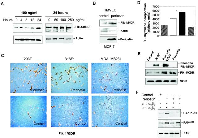
FIG. 4.
Up-regulation of Flk-1/KDR is at least in part responsible for periostin-induced angiogenesis. (A) Recombinant periostin induces the expression of Flk-1/KDR in a time- and dose-dependent manner. HMVEC were treated with periostin (100 ng/ml) for up to 24 h or treated with different concentrations of periostin for 24 h. Protein samples from the treated cells were analyzed for Flk-1/KDR expression by immunoblotting. Actin was used as a loading control. (B) Periostin derived from conditioned medium of MCF-7 cells induces Flk-1/KDR expression. Control or periostin-producing MCF-7 cells shown at the bottom panel were incubated with serum-free medium for 48 h. HMVEC were incubated with the conditioned media for 24 h prior to analysis for Flk-1/KDR expression. (C) Increased presence of Flk-1/KDR associated with blood vessels is detected in tumors derived from periostin-producing cells. Tumor sections derived from 293T, B16F1, or MDA-MB-231 cells were subjected to immunohistochemical staining analysis with an anti-Flk-1/KDR antibody. (D) Periostin pretreatment potentiates HMVEC proliferation in response to VEGF. HMVEC were pretreated with periostin (100 ng/ml) for 12 h. After the cells were washed, VEGF (10 ng/ml) was added for 12 h to determine the effect on cell proliferation. *, P was <0.05 compared with VEGF treatment alone. (E) Periostin pretreatment potentiates Flk-1/KDR phosphorylation in response to VEGF. The same assay condition was used as described above except that the incubation time for VEGF was 5 min, followed by the quantitative determination of phosphorylated Flk-1/KDR by immunoblotting with a specific anti-phospho-Tyr 951 antibody.

