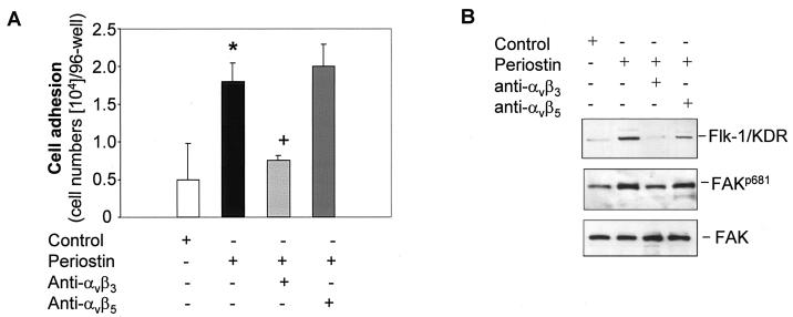
FIG. 5.
The αvβ3 integrin-FAK signaling pathway mediates the induction of Flk-1/KDR expression. (A) HMVEC (5 × 10⁴ cells/well) were cultured in a 96-well plate precoated with periostin (10 μg/ml) in the presence of anti-αvβ3 or anti-αvβ5 integrin antibody (10 μg/ml) for 1 h (noncoated wells were used as the control). Adhesive cells were counted following washing. *, P was <0.05 compared with the control cells; +, P was <0.05 compared with the cells plated on periostin-coated wells alone. (B) in the presence or absence of the indicated specific anti-integrin antibodies, HMVEC were treated with periostin (100 ng/ml) for 12 h to examine the effect on the up-regulation of Flk-1/KDR as determined by Western blotting. Alternatively, the same assay conditions were used except that the time of incubation was reduced to 15 min to observe a change in FAK autophosphorylation on Tyr 681 (FAKp681). An antibody against FAK was used as a loading control.

