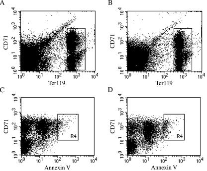
FIG. 5.
Erythroid cell distribution and apoptosis in bone marrow of Rps19+/− mice. The fluorescence-activated cell sorting analysis data show cells of the erythroid lineage from adult mouse bone marrow cells of one Rps19+/− male (A and C) and one Rps19+/+ male control (B and D). CD71+/Ter119high cell populations (boxed areas in panels A and B) were gated and analyzed for apoptosis by using annexin V as the marker (C and D). An area (R4 boxes) of increased annexin V binding was defined as containing apoptotic cells and included 1.9% of the total CD71+/Ter119high cells for the Rps19+/− mouse and 3.1% for the wild-type control.

