Figure 1.
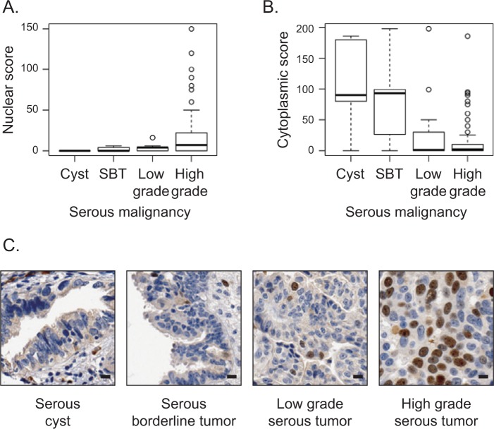
Nuclear localization of Ect2 correlates with advanced disease in human serous epithelial ovarian cancers. Ect2 subcellular localization was analyzed by immunohistochemistry (IHC) using a well-validated26-29 ovarian tissue microarray (TMA). Tumor cores were stained with Ect2 antibody (1:350; Millipore, Billerica, MA) and scored for percentage cells stained as well as intensity of staining in each location. Separate nuclear scores and cytoplasmic scores were calculated as described in Materials and Methods and binned according to disease severity. Quantification of Ect2 localization showed (A) significantly greater nuclear expression (P = 0.0001516) and (B) significantly lower cytoplasmic expression (P = 0.0007163) in advanced disease. SBT = serous borderline tumor. Box and whisker plots include horizontal lines corresponding to median, first and third quartiles (hinges), and extreme points excluding outliers (whiskers). (C) Representative images of a benign serous cyst, a serous borderline tumor, a low-grade and a high-grade serous epithelial tumor stained for Ect2 illustrate increased nuclear staining upon disease progression. Scale bars represent 10 µm.

