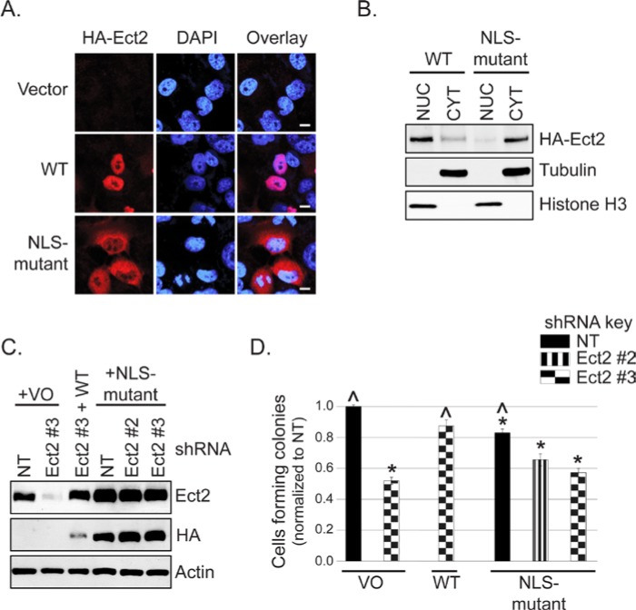
Figure 5.
Predominantly nuclear localization is required for Ect2-mediated transformed growth. (A) Disruption of the nuclear localization signals (NLSs) of Ect2 impairs its nuclear localization compared with WT Ect2. Localization of an Ect2 NLS-mutant (R348,349,350,370,372A) in which both NLSs have been disrupted was determined by confocal immunofluorescence microscopy, probing for HA-tagged Ect2 (red, 1:500, clone 16B12, Covance, Princeton, NJ) and the nuclear marker DAPI (blue), n = 3. Scale bars represent 10 µm. (B) NLS-mutant Ect2 is localized predominantly in the cytoplasm. The shift in localization of Ect2 on NLS mutation was confirmed by fractionating cells expressing either NLS-mutant or WT Ect2, and blotting for HA-tagged Ect2 (1:1,000, Covance). Purity of each fraction was confirmed via immunoblot using anti-histone H3 antibody (1:12,000, Ab1791, AbCam, Cambridge, MA) as a nuclear marker and tubulin as a cytoplasmic marker, with equal amounts of the nuclear and cytoplasmic fractions loaded onto the gel. Average percentage of Ect2 in the nucleus was calculated from densitometry values. Whereas 72.3% of WT Ect2 was observed in the nucleus, only 10.4% of NLS-mutant Ect2 was nuclear (n = 2). (C) NLS-mutant Ect2 restored Ect2 expression in knockdown cells to a higher degree than WT. The shRNA-resistant mutants indicated above were stably expressed in Ect2-knockdown OVCAR8 cells. Cell lysates were immunoblotted as in Figure 4B except that actin served as a loading control. (D) Ect2 NLS-mutant failed to rescue colony formation in soft agar. WT but not NLS-mutant Ect2 rescued the impairment of soft agar colony formation caused by Ect2 knockdown (n = 5). Error bars represent SEM. Statistically significant differences are marked with * and ^ as in Figure 4C.

