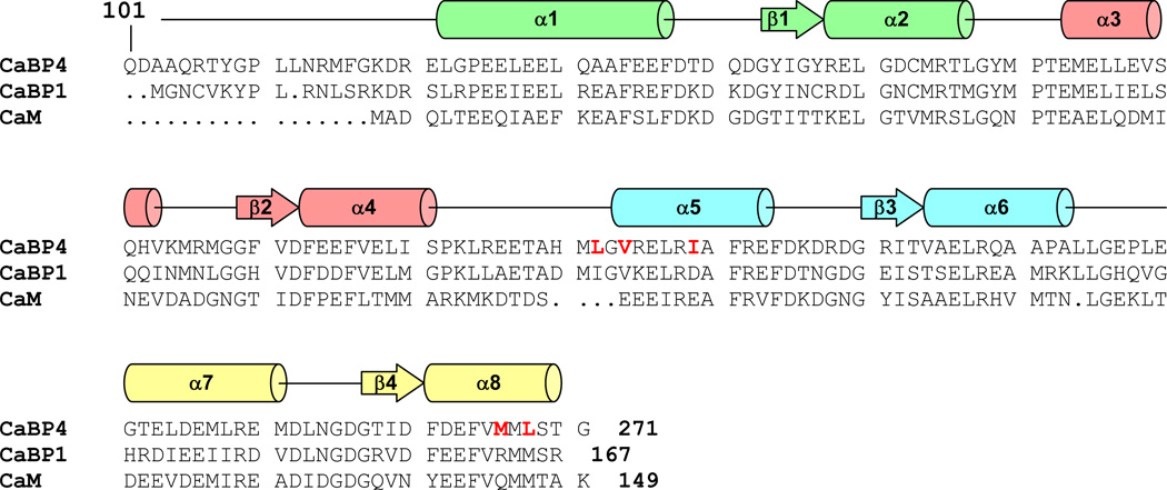
Figure 1.
Alignment of the primary sequence of mouse CaBP4, CaBP1, and calmodulin. Secondary structural elements indicated schematically were derived from analysis of NMR chemical shift index (CSI) (Wishart et al., 1992). The four EF-hands (EF1, EF2, EF3 and EF4) are highlighted green, salmon, cyan, and yellow, respectively. CaBP4 residues highlighted in red exhibit markedly different backbone chemical shift values compared to those of CaBP1 (Li et al., 2009).

