FIG. 2.
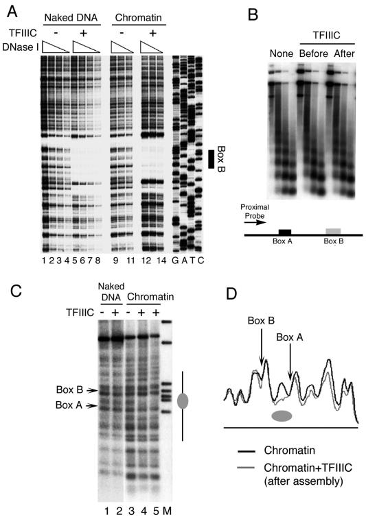
Chromatin structure and TFIIIC binding. (A) DNase I footprinting of pCS6 as naked DNA (lanes 1 to 8) or assembled into chromatin (lanes 9 to 14) was performed in the absence (lanes 1 to 4 and 9 to 11) or presence (lanes 5 to 8 and 12 to 14) of TFIIIC. Box B is identified on the right. The 5′ end of the primer extension probe was 135 bp away from the 3′ end of box B, complementary to the U6 gene top strand. (B) TFIIIC does not disrupt the periodicity of nucleosomal arrays. MNase-digested chromatin fragments, from assemblies in the absence or presence of TFIIIC added at different times, were purified, electrophoretically resolved, blotted, and hybridized to a probe complementary to DNA ∼50 bp upstream of the start site (proximal probe), as shown schematically at the bottom of the panel. (C) IEL analysis of chromatin structure on the U6 snRNA gene. Naked DNA (lanes 1 and 2) and chromatin (lanes 3 to 5) were digested with MNase and probed with a primer that hybridizes 894 bp upstream of the SNR6 TATA box. Lane M represents markers, lane 3 is the control chromatin sample, and lanes 4 and 5 had TFIIIC added prior to and after 4.5 h of chromatin assembly, respectively. Positions of boxes A and B are marked on the left. The gray ellipse on the right marks the nucleosome-size protection between boxes A and B. (D) Aligned profiles of lanes 5 and 3 of panel C. The gray ellipse marks the nucleosomal protection between boxes A and B.

