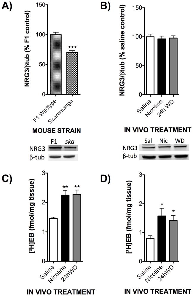
Figure 4. Chronic treatment with nicotine and withdrawal results in differential biochemical responses in the F1 wildtype and NRG3ska mice.
F1 hybrid and NRG3ska mice were treated in vivo with either saline, nicotine (18 mg/kg/day), or undergoing 24h withdrawal (WD) from nicotine. A) Hippocampal tissues from saline treated animals were used in Western blot analysis to evaluate basal levels of NRG3 in the two strains of mice. NRG3ska mice demonstrated a signficant reduction in NRG3 levels compared to F1 hybrids (N=8). Representative blots are shown below. B) Hippocampal tissues from NRG3ska mice treated with saline, nicotine, or 24h WD were used in Western blot analysis to evaluate treatment changes in NRG3 levels. NRG3ska mice did not show an induction of NRG3 following chronic nicotine or 24h WD (N=8). Representative blots are shown below. C) Hippocampal tissues from F1 hybrid mice treated with saline, nicotine, or 24h WD were used for homogenate-binding experiments with a saturating concentration of [3H]epibatidine ([3H]EB, 1.5 nM). F1 hybrid mice chronically treated with either nicotine or 24h WD displayed an increased density of nAChRs compared to saline controls (N=4). D) Hippocampal tissues from NRG3ska mice treated with saline, nicotine, or 24h WD were used for homogenate-binding experiments with a saturating concentration of [3H]epibatidine ([3H]EB, 1.5 nM). NRG3ska mice chronically treated with either nicotine or 24h WD displayed an increased density of nAChRs compared to saline controls (N=4). *, p<0.05; **, p<0.01; ***, p<0.005.

