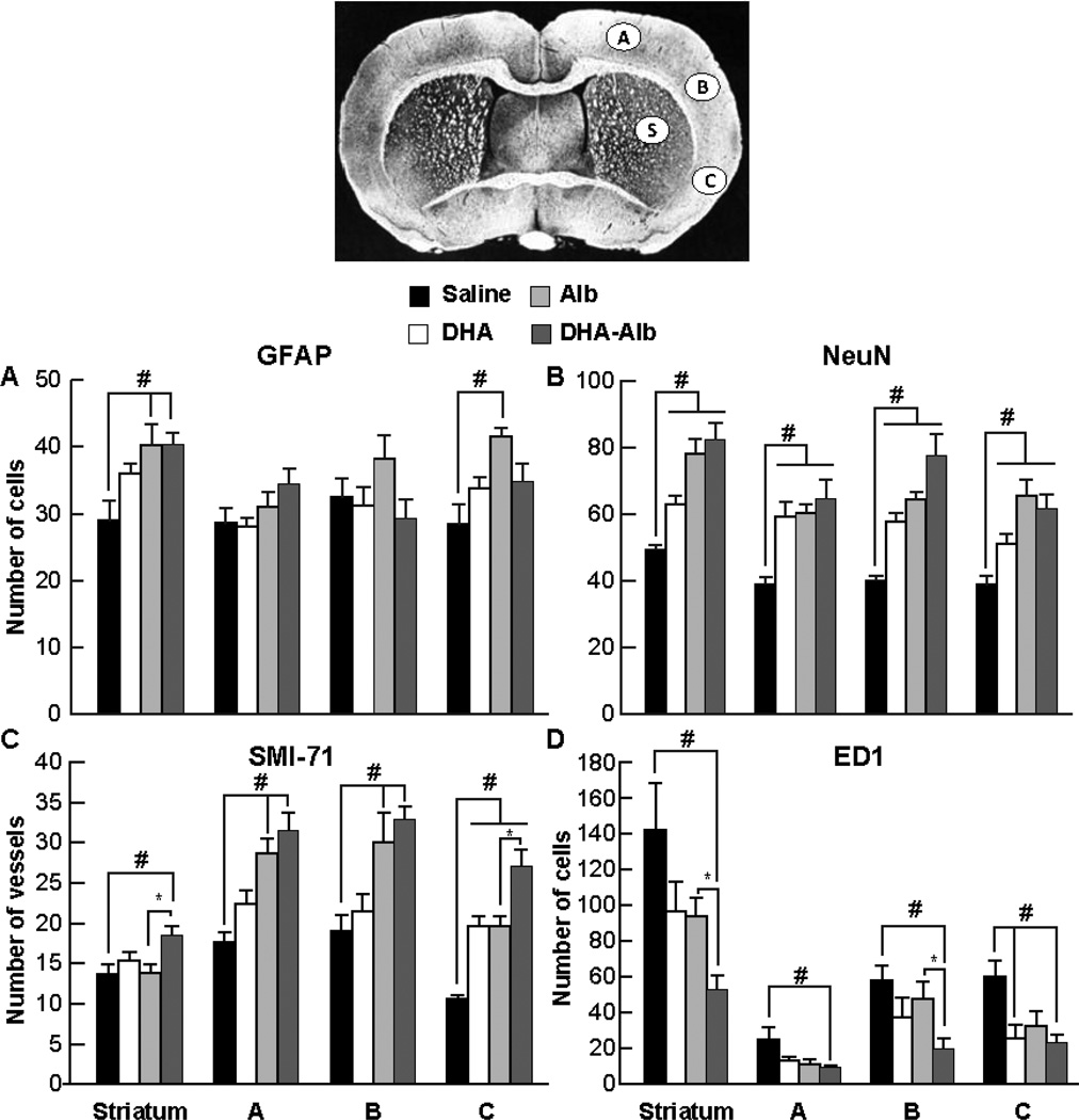
Figure 4.
Cell count for GFAP positive astrocytes, NeuN positive neurons, SMI-71 positive vessels and ED1 positive microglia cells on day 7 after 2h of MCAo. Coronal brain diagram showing locations of regions for cell count (bregma level - 0.3 mm) in cortex (A, B, and C) and striatum (S). Alb- and DHA-Alb-treated rats expressed increased numbers of GFAP compared to the saline-treated group (Panel A). All treatments (DHA, Alb, DHA-Alb) increased numbers of NeuN positive cells and SMI-71 immunopositive vessels compared to saline-treated rats (Panels B and C). In addition, DHA-Alb treatment increased SMI-71 positive vessels compared to the corresponding Alb group (Panel C). DHA, Alb, and DHA-Alb treatments decreased microglial invasion of the infarcted tissue while salinetreated rats showed significant microglial infiltration of the infarct regions (Panel D). In addition, DHA-Alb treatment decreased ED1 positive microglia compared to the corresponding Alb group (Panel D). Values shown are means ± SEM., *P<0.05 #P<0.05 versus saline group; *P<0.05 DHA-Alb versus Alb group (repeated measures ANOVA followed by Bonferroni tests).

