
Figure 2. Histone H4 is decreased upon neutrophil depletion.
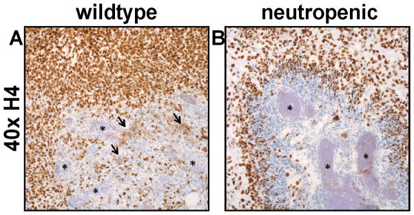
(A and B) Immunohistochemistry for histone H4. (A) In the SRF3-treated mouse (wildtype), there is increased extracellular immunoreactivity for histones (arrows) in proximity to the bacterial cells (*). (B) Anti-Gr1(RB6)-treated mice (neutropenic) have fewer infiltrating cells, very little extracellular histone H4, and large bacterial aggregates (*).