FIGURE 9.
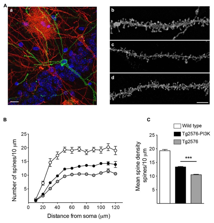
PI3K increases spine density in a Tg2576 mice model. (A) (a) Example of biolistic stain, the image shows an area from the basal CA1 region of a 6-month-old Tg2576 mouse.Three hippocampal neurons appear stained with DiI (red) or DiO (green). The stratum pyramidale nuclei appear in blue (DAPI). An astrocyte, red area in the top middle side of the picture was also labeled. Scale bar: 10 μm (a). Representative dendritic segments for Wild-type (b), Tg2576 (c), and Tg2576 injected with PTD4-PI3KAc (d). Scale bar = 3 μm. (B) Number of spines along basal dendrites in neurons from wild-type (open circles), Tg2576 (gray circles), and PTD4-PI3KAc-treated Tg2576 mice (closed circles). The values were statistically different between 30 and 120 μm from the soma (two-way ANOVA, p < 0.001; significant levels are not included for clarity). (C) Mean spine densities considering all values 60 μm or more away from the soma (a distance at which the number of spines have reached a plateau). The number of spines in Tg2576 mice (10.5 ± 0.16, 50 dendrites from four animals) is significantly reduced compared to the wild-type (19.3 ± 0.47, 30 dendrites from three mice). Tg2576 injected with PTD4-PI3KAc exhibited an intermediate value (13.8 ± 0.18, 50 dendrites from six animals), suggesting that overactivation of PI3K partially counteracts the loss of spines in Tg2576 mice.

