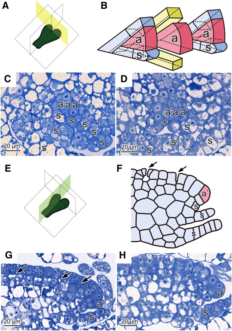
Figure 2.
Histological Observation of Thalli of the Wild Type and nop1 Mutant.
(A) Diagram showing transverse planes in which thallus sections were cut.
(B) Schematic illustration of the pattern of dorsiventral division of the apical cell and subapical cells of M. polymorpha.
(C) and (D) Transverse sections of wild-type (C) and nop1 (D) thalli.
(E) Diagram showing vertical longitudinal planes in which thallus sections were cut.
(F) Schematic illustration of a typical vertical longitudinal section around the subapical cell of wild-type M. polymorpha.
(G) and (H) Vertical longitudinal sections of wild-type (G) and nop1 (H) thalli.
Arrows indicate the initiation of ICSs in the wild type ([F] and [G]). a, apical cell; a’, subapical cell; s, ventral scale.

