Figure 6.
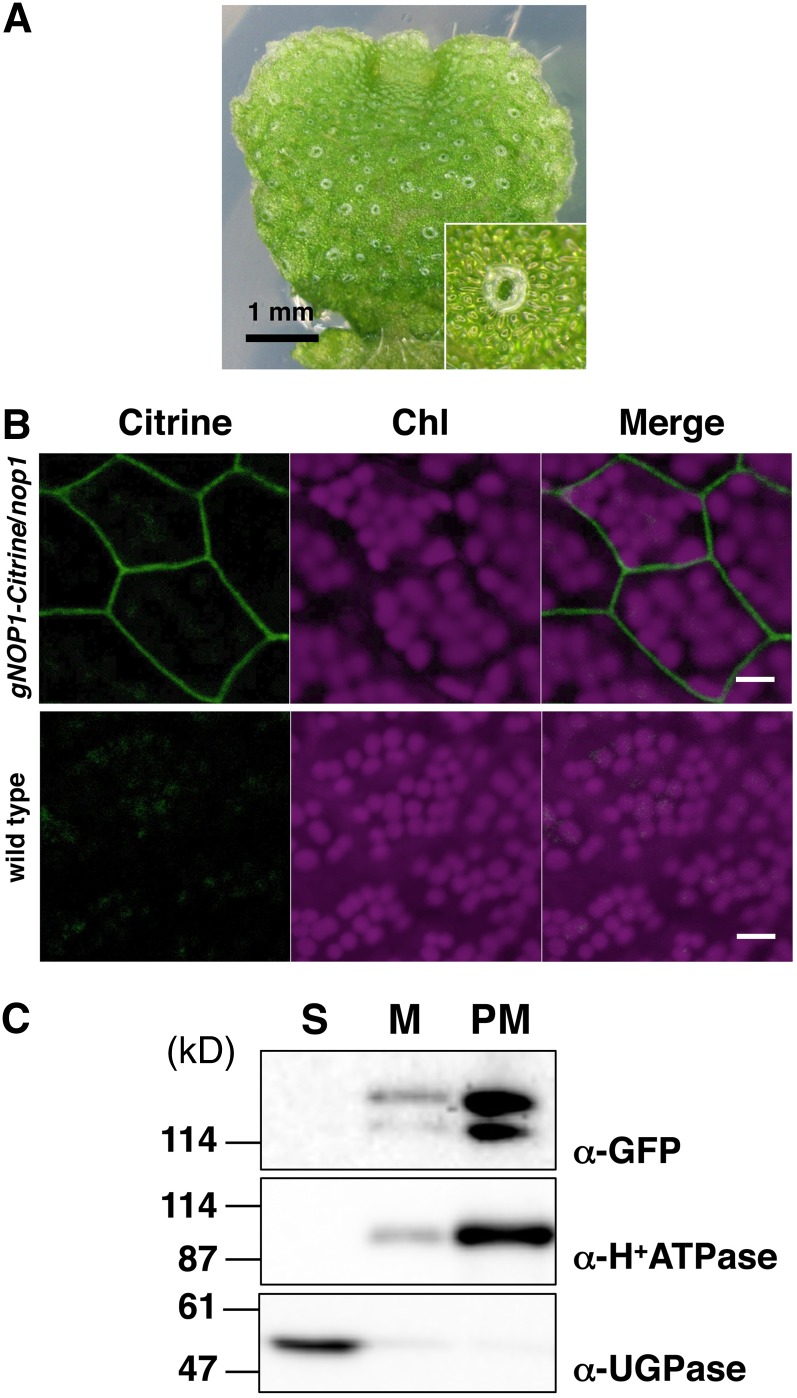
Plasma Membrane Localization of NOP1.
(A) Phenotype of gNOP1-Citrine/nop1 #1 and a magnified image (inset). Introduction of the NOP1 gene fused with Citrine in the C terminus rescued the impaired air-chamber phenotype of nop1.
(B) Confocal microscopy of thallus cells. The panels show Citrine fluorescence, chlorophyll (Chl) autofluorescence, and merged images from left to right, respectively. Thallus cells of the gNOP1-Citrine/nop1 #1 (top panels) and the wild type (bottom panels) were observed. Bar = 10 µm.
(C) Protein gel blot analysis of the soluble fraction (S), total membrane fraction (M), and plasma membrane fraction (PM) from gNOP1-Citrine/nop1 #1. Fusion proteins were detected with an anti-GFP antibody. H+-ATPase and UGPase were chosen as subcellular markers for the plasma membrane–bound protein and for the cytosolic protein, respectively.

