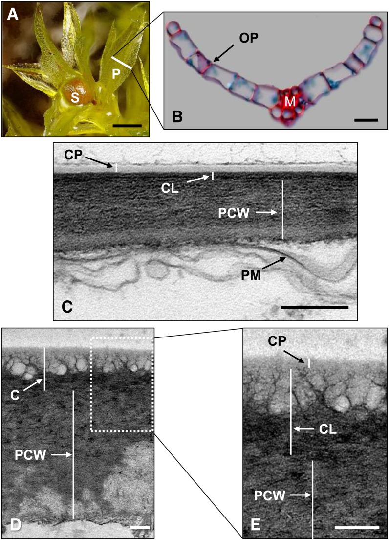
Figure 1.
P. patens Morphology and Cuticle Structure.
(A) P. patens gametophore with several phyllids (P) and an immature sporophyte (S) growing from the apex.
(B) P. patens phyllid shown in transverse section, corresponding to the white line in (A), consisting of a single-cell-layer-thick lamina attached to a central midrib (M). Cell walls are stained red and intracellular contents blue. OP, outer periclinal wall.
(C) Transmission electron micrograph of the phyllid outer periclinal cell wall, showing the layers of the cuticle (CL), polysaccharide cell wall (PCW), and plasma membrane (PM).
(D) Transmission electron micrograph of the sporophyte capsule outer periclinal cell wall (cuticle [C]).
(E) Expanded view of the box in (D).
Bars = 750 μm in (A), 20 μm in (B), and 200 nm in (C) to (E).

