
Figure 8.
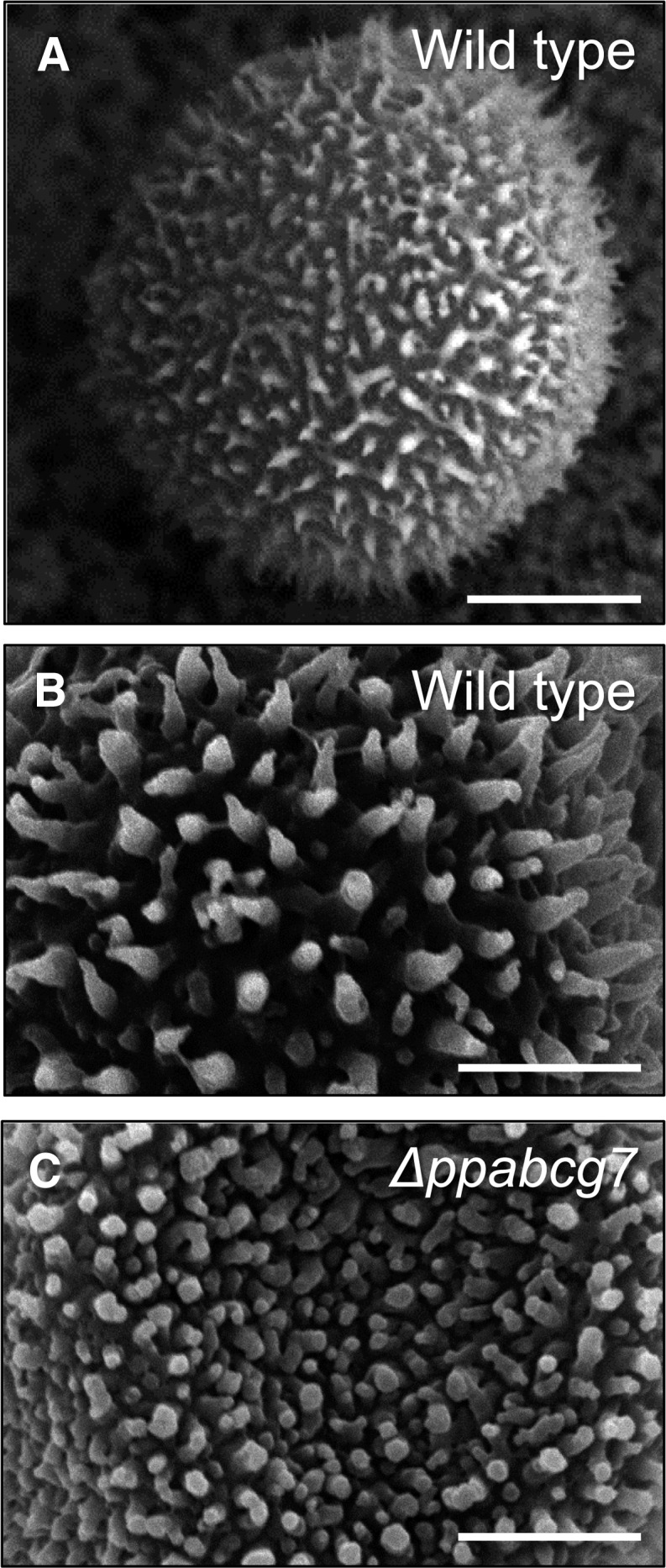
P. patens Spore Phenotypic Analysis.
Scanning electron micrographs of spore surface decorations in the wild type ([A] and [B]) and Δppabcg7 (C), showing irregular and rounded protrusions in the mutant. Bars = 10 μm in (A) and 5 μm in (B) and (C).